Ct Scan Brain Made Easy Pdf
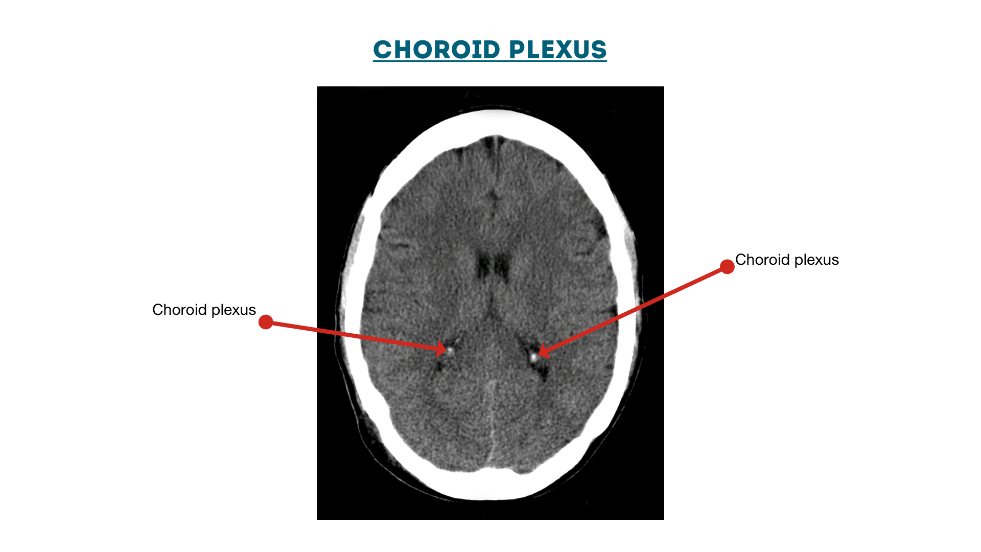
Ct scan brain made easy pdf. Shift - the falx should be in the midline with ventricles the same on both sides. Hartmut Gross at the Rural Emergenc. Symmetry - make sure sulci and gyri appear the same on both sides.
Introduction CT anatomy abdomen CT section with pathology Take home message 6. The introduction of increasingly sophisticated imaging techniques over the last few decades has transformed neurological diagnosis. Isodense with brain 2-3wks.
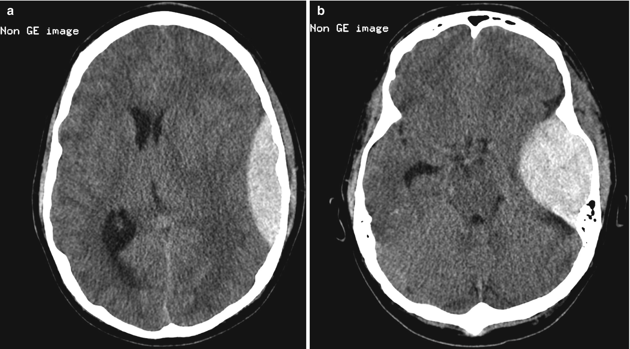
Intraparenchymal HemorrhageContusions Sudden deceleration of the head causes the brain to impact on bony prominences eg temporal frontal occipital poles. We would like to show you a description here but the site wont allow us. Ischaemic stroke tumour or cerebral abscess.
This is a lecture given to emergency medicine providers discussing how to read a head CT. On a normal CT head scan the grey and white matter should be clearly differentiated. Abdominal CT scan made easy 1.
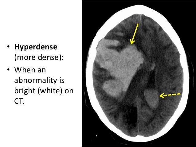
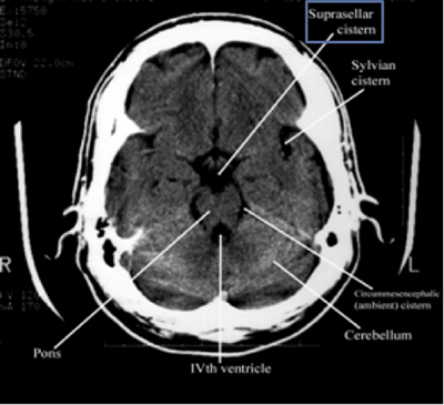
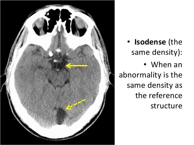
Blood will 1st become isodense with the brain 4 days to 2 weeks depending on clot size and finally darker than brain 2-3 weeksii C Cisterns Cerebrospinal fluid collections jacketing the brain. A cranial CT scan is known by a variety of names as well including brain scan head scan skull scan. CT BRAIN SCAN Mamdouh Mahfouz MD Professor of Radiology Cairo University Author.
A neurologist needs to be able to request the appropriate testmay need to consult with radiological colleagues in unusual casesand know what. Compare side to side. It was given on 92615 by Dr.
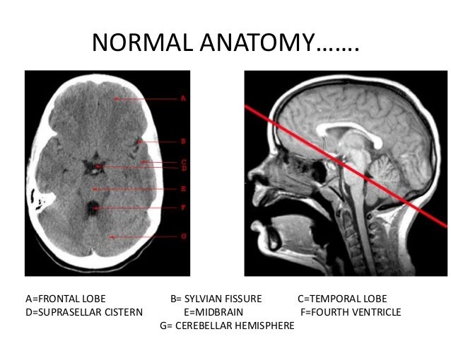
SCHEME OF THE LECTURE BASIC PRINCIPLES OF CT SCAN NORMAL NEUROANATOMY AS SEEN ON HEAD CT SCANS ILLUSTRATIONS 4. Non-traumatic hemorrhagic lesions seen more frequently in elderly and located in basal ganglia.
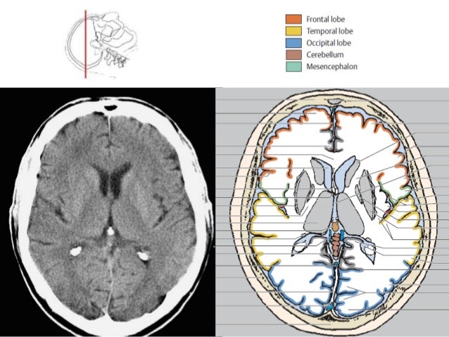
Head CT Approach First - evaluate normal anatomical structures window for optimal brain tissue contrast Second assess for signs of underlying pathology such as.
Shift - the falx should be in the midline with ventricles the same on both sides. Download Clinical radiology made ridiculously simple pdf February 9 2016 by Dr Hamza Arshad 5 Comments Radiology is a branch of medial sciences that deals with the diagnosis and treatment of diseases with radiations. SCHEME OF THE LECTURE BASIC PRINCIPLES OF CT SCAN NORMAL NEUROANATOMY AS SEEN ON HEAD CT SCANS ILLUSTRATIONS 4. Hypoperfused brain at risk for progression to infarction salvageable Usually located around the ischemic core and represents target of reperfusion therapy Is represented by total area of hypoperfused brain MINUS infarcted core Depicted by prolonged time it takes for contrast to reach and traverse areas of the brain. Hypodense with brain TypesLocations. Intraparenchymal HemorrhageContusions Sudden deceleration of the head causes the brain to impact on bony prominences eg temporal frontal occipital poles. Head CT Approach First - evaluate normal anatomical structures window for optimal brain tissue contrast Second assess for signs of underlying pathology such as. Symmetry - make sure sulci and gyri appear the same on both sides. Shift - the falx should be in the midline with ventricles the same on both sides.
In this article we will outline the basic science behind CT scans describe the principles of interpretation and highlight their advantages and drawbacks compared to other imaging techniques. Non-traumatic hemorrhagic lesions seen more frequently in elderly and located in basal ganglia. Download Clinical radiology made ridiculously simple pdf February 9 2016 by Dr Hamza Arshad 5 Comments Radiology is a branch of medial sciences that deals with the diagnosis and treatment of diseases with radiations. In this article we will outline the basic science behind CT scans describe the principles of interpretation and highlight their advantages and drawbacks compared to other imaging techniques. 542014 121052 AM. Hypodense with brain TypesLocations. Isodense with brain 2-3wks.















Posting Komentar untuk "Ct Scan Brain Made Easy Pdf"